


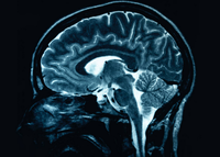
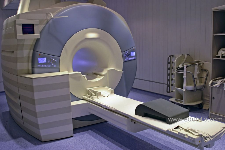
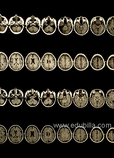
Magnetic resonance imaging or scanning (also called an MRI) is a method of looking inside the body without using surgery, harmful dyes or x-rays. The MRI scanner uses magnetism and radio waves to produce clear pictures of the human anatomy.
Damadian's early work on NMR concerned investigating potassium ions inside cells.He found that the potassium relaxation times were much shorter compared with aqueous solutions of potassium ions. This suggested that potassium was not free but complexed to ‘fixed-charge’ counter-ions, as he had previously determined.
He and other researchers independently investigated the signals of 1H NMR in cells, and found that the relaxation times were much shorter than in distilled water. This was consistent with ordering of a large part of the water by adsorption onto macromolecular surfaces. Damadian predicted that cancerous cells would have longer relaxation times, both because of the disordering of malignant cells and because of their elevated potassium levels, since the potassium ions would be ‘structure-breaking’ to the ordered water fraction.
In a 1971 paper in the journal Science, SUNY Downstate Medical Center professor Damadian reported that tumors can be detected in vivo by nuclear magnetic resonance (NMR) because of much longer relaxation times than normal tissue. He suggested that these differences could be used to detect cancer, even in the early stages where it would be most treatable, though later research would find that these differences, while real, are too variable for diagnostic purposes. However, Damadian in his seminal paper claimed only that his method was a detection tool, making no claim about being a diagnostic tool, but intended that it would provide a non-invasive way of detecting cancers and monitoring the effectiveness of their therapy.
According to a Wall Street Journal article, Damadian's initial methods were flawed for practical use, relying on a point-by-point scan of the entire body and using relaxation rates, which turned out to not be an effective indicator of cancerous tissue. However, the same article pointed out, “Nevertheless, his observation of T1 and T2 differences in cancerous tissue was a Eureka moment for Paul Lauterbur.” Furthermore, Damadian's seminal paper documented in its Table 2 that T1 relaxation times were different, beyond experimental uncertainty, across all his samples over different healthy tissues: rectus muscle, liver, stomach, small intestine, kidney, and brain. This showed the way to accurate imaging of the body's soft tissues for the first time; X-ray imaging was severely deficient for soft tissue analysis because the difference in absorption was so small (<4%). So when in the court case Fonar v. General Electric, GE's attorneys made the same claim that relaxations times were also prolonged in non-cancerous tissue, so were not a good diagnostic, Fonar's attorneys responded that it was unfair to punish Damadian because his methods detected even more features than he had intended. Indeed, even today, 90% of MRI scans on patients produce images that are relaxation dependent, either T1- or T2-dependent images.
In 1974, he received the first patent in the field of MRI when he patented the concept of NMR for detecting cancer after filing an application in 1972—now there are over 4,500 patents on MRI. As the National Science Foundation notes, “The patent included the idea of using NMR to ‘scan’ the human body to locate cancerous tissue.”However, it did not describe a method for generating pictures from such a scan or precisely how such a scan might be done,.But without Damadian's discovery of the profound sensitivity of the relaxation time to different tissue types and malignant tissue, there would be no picture at all.
In the 1950s, Herman Carr reported creating a one-dimensional MR image. Prompted by Damadian's report on the potential medical uses of NMR, Paul Lauterbur expanded on Carr's technique and developed a way to generate the first MRI images, in 2D and 3D, using gradients. Peter Mansfield from the University of Nottingham then developed a mathematical technique that would allow scans to take seconds rather than hours and produce clearer images than Lauterbur had. While Lauterbur and Mansfield focused on animals and human limbs, Damadian built the first full-body MRI machine and produced the first full magnetic resonance imaging ("MRI") scan of the human body, albeit using a “focused field” technique that differs considerably from modern imaging. In recording the history of MRI, Mattson and Simon (1996) credit Damadian with describing the concept of whole-body NMR scanning, as well as discovering the NMR tissue relaxation differences that made this feasible.


")


