


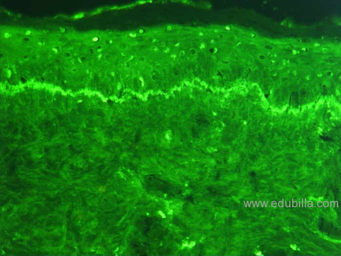
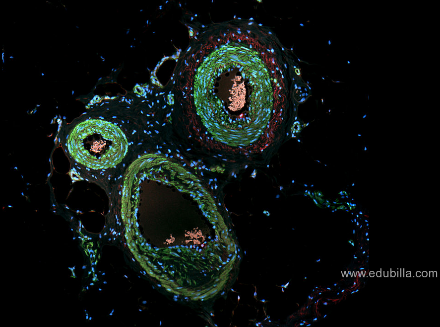
Immunofluorescence is a technique used for light microscopy with a fluorescence microscope and is used primarily on microbiological samples. This technique uses the specificity of antibodies to their antigen to target fluorescent dyes to specific biomolecule targets within a cell, and therefore allows visualisation of the distribution of the target molecule through the sample. Immunofluorescence is a widely used example of immunostaining and is a specific example of immunohistochemistry that makes use of fluorophores to visualise the location of the antibodies.
This method of study focuses on the immune response that occurs within a diseased tissue or its cells, a behavior researchers can observe after they apply a fluorescent stain either directly or indirectly to the specimen.
In studying these cells and their reactions, scientists are able to determine which specific proteins protect the body against those particular foreigners that invade it with the intent of causing sickness or in some cases, death.
Applications
Scientists can use the immunofluorescence technique to perform a variety of lab tests and observations, each of which provides a fresh perspective for the sample currently undergoing analysis.
For instance, a researcher may opt to use this testing method to examine tissue samples and beads, detect particular proteins using microarrays, or they might choose to evaluate cultured cells, as well as those in suspension.
An observer can apply this technique to samples that are fixed or fresh, thus providing more opportunity for diverse analysis and accurate results.
A researcher may also choose to apply this technique when studying DNA sequences on chromosomes, as these particles are extremely small and hard to detect. Obtaining spatial data concerning tissue or cell genetics is another way to use immunofluorescence, as is the observation of parasites and bacteria.
The deeper layers of a cell or tissue, along with the antigens that infect these layers, is another area that scientists can study using this staining method.


")


