Golgi's method is a silver staining technique discovered by Italian physician and scientist Camillo Golgi (1843–1926) in 1873 that is used to visualize nervous tissue under light microscopy. It was initially named the black reaction (la reazione nera) by Golgi, but it became better known as the Golgi stain or later, Golgi method.
In contrast, the Golgi method, focused in this review, has allowed for the visualisation of entire neurons and glia in high detail and with good contrast. Moreover, compared to Nissl staining and silver impregnation, the Golgi method has the beneficial feature of characteristically selective staining. Because neurons are stained only sparsely with the Golgi method, it is a powerful staining technique for providing a complete, detailed representation of a single neuron. The aim of this review was to discuss the history and evolution of the Golgi method.
Mechanism of Golgi method
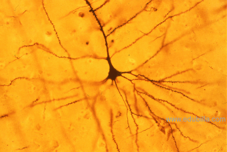
In the Golgi method, it is possible to stain various neural cells such as neurons and neuroglia, as well as cells from the vascular system. The crystal of the silver chromate taken up by neurons spreads into every corner of the neuron. Moreover, the Golgi method provides images of neurons as dark features on a transparent background with good contrast. Therefore, the method enabled the discovery of previously unknown neuronal structures such as growth cones, dendritic spines and filopodia.
As mentioned above, the Golgi method has an exclusive property – its capricious selectivity. As a potential mechanism underlying such selectivity, Shankaranarayana et al.speculated that the stainability of an individual cell might be significantly affected by interaction between the cellular status during fixation (e.g. metabolic state and pH) and the heavy metal used for staining. We have recently acquired evidence to support this assumption. Our report demonstrated that a modified Golgi method used for staining primary cultured neurons did not possess selectivity, typical of the Golgi technique. The underlying reason was believed to be that the bioactivity of each cultured neuron was extremely uniform due to the re-initialisation of the hippocampal neuronal cells after harvesting in the culture medium. However, the precise mechanism of selective stainability remains to be confirmed. The unique selective stainability of the Golgi method has allowed visualisation of the entire morphology of single neurons. This has enabled researchers to determine the projection sites as well as the morphology of neuronal cells.
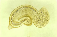
Traditional methods of analysis applied to Golgi-stained samples included observational study using light microscopy or measurements provided by camera lucid drawings. A great deal of information is available from analysing such photos and images, including the total length of neuronal processes, number of branch points, complexity of the dendritic tree and types of dendritic spines. Further, recent improvements in microscopy and computer software have allowed high-quality images to be obtained and analysed in three dimensions. The accumulation of such information has led not only to the revelation of detailed morphological aspects of neural circuitry in various parts of the brain, but also to information on abnormal neuronal morphologies encountered in brains with neuronal diseases.