Electroencephalography is the neurophysiologic measurement of the electrical activity of the brain by recording from electrodes placed to the scalp, or in the special cases on the cortex. The resulting traces are known as an electroencephalogram (EEG) and represent so-called brainwaves. This device is used to assess brain damage, epilepsy and other problems. In some jurisdictions it is used to assess brain death. EEG can also be used in conjunction with other types of brain imaging.
Neuroscientists and biological psychiatrists use EEGs to study the function of the brain by recording brainwaves during controlled behavior of human volunteers and animals in lab experiments. Theories to explain sleep often rely on EEG patterns recorded during sleep sessions. In addition, the procedure is used clinically to assist in the diagnosis of epilepsy.
The first EEG recording, obtained by Hans Berger in 1929.
Methods
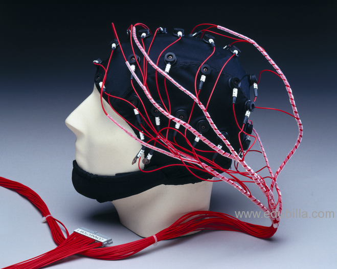
The recording is obtained by placing electrodes on the scalp, usually after preparing the scalp area by light abrasion and application of a conductive gel to reduce impedance. Each electrode is connected to an input of a differential amplifier (one amplifier per pair of electrodes), which amplifies the voltage between them (typically 1,000–100,000 times, or 60–100 dB of voltage gain), and then displays it on a screen or inputs it to a computer. The amplitude of the EEG is about 100 µV when measured on the scalp, and about 1-2 mV when measured on the surface of the brain.
EEG has several limitations. Scalp electrodes are not sensitive enough to pick out individual action potentials, the electric unit of signaling in the brain, or whether the resulting electrical activity is releasing inhibitory, excitatory or modulatory neurotransmitters. Instead, the EEG picks up synchronization of neurons, which produces a greater voltage than the firing of an individual neuron. Secondly, EEG has limited anatomical specificity when compared with other functional brain imaging techniques such as functional magnetic resonance imaging (fMRI). Some anatomical specificity can be gained with the use of EEG topography, which uses a large number of electrodes to triangulate the source of the electrical activity.
EEG has several strong sides as a tool of exploring the brain activity. The time resolution is very high. As other methods for researching brain activity have time resoloution between seconds and minutes, the EEG has a resolution down to sub-millisecond. The brain is thought to work through its electric activity. EEG is the only method to measure it directly. Other methods for exploring functions in the brain do rely on blood flow or metabolism which may be decoupled from the brain electric activity. Newer research typically combines EEG or MEG with MRI or PET to get high temporal and spatial resolution.